Microscopy
Microscopy
The Microscopy Core comprises many newer, advanced microscopes including widefield, laser scanning confocal, spinning disk confocal, multiphoton, and semi-super-resolution microscopes which are suitable for fixed samples, live-cell, and intravital experiments. Additionally, the VBRI has several formidable analysis stations running up-to-date versions of Nikon NIS-Elements and Imaris. Experienced Imaging Specialists are available for consultations regarding experimental design and sample preparation, as well as provide training in microscope use, image acquisition, and data analysis.
Instrumentation and Services
3I Spinning Disk Confocal Microscope
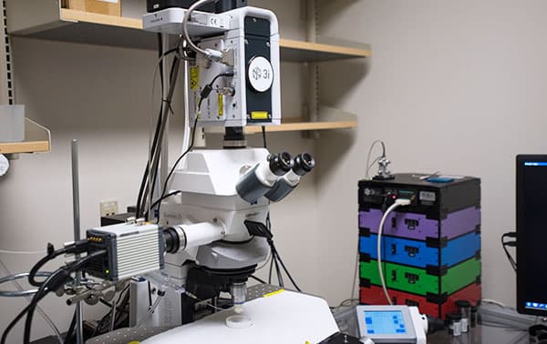
The spinning disk confocal microscope is specifically designed to enable rapid, real-time, in-vivo studies of thrombosis and hemostasis in small animals. The system consists of an automated Yokogawa spinning disk confocal unit; a high-speed, high-sensitivity camera; four solid-state excitation lasers; and an ablation laser for creating thrombi via laser injury. This upright microscope can accommodate small animals and stereotaxic equipment under its vertical illuminator. This instrument is operated by Microscopic Imaging Core staff. For consultation, contact mschulte@versiti.org or request consultation below.
Nikon Widefield
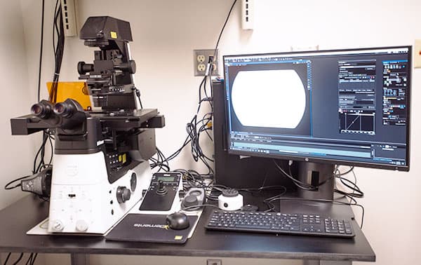
The Nikon Ti2 Widefield inverted microscope is a high-speed, motorized microscope for advanced imaging. This microscope is primarily used for experiments with multidimensional imaging—it can combine multi-channel, multi-XY positions, Z-stacking, and large image stitch collection. The large field-of-view is optimized for collecting excellent large image stitches using fluorescence and/or brightfield. It has a dual-camera system for fluorescence (ORCA Fusion CMOS) or RGB color capture for non-fluorescent stains (DS-Ri2; 16.25 MP). The fluorescence light source is a Lumencor SPECTRA III LED light engine (390/22, 440/20, 475/28, 510/25, 555/28, 575/25, 637/12, 748/12). The objectives (10x, 20x, 40x, 60x Oil, 100x Oil) are designed to image through the thickness of a “No. 1.5” glass coverslip.
Nikon Spinning Disk Confocal +SoRa
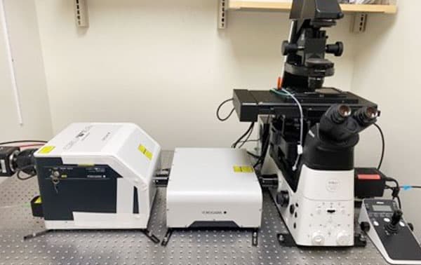
A Nikon Ti2 inverted microscope is equipped with the Yokogawa CSU-W1 SoRa, a confocal field scanning spinning disk system with super-resolution by optical reassignment. Primarily used for live-cell imaging at high speed, super resolution, and colocalization studies in live or fixed cells. This system is great for multidimensional imaging that combines multi-channel, multi-XY positions, Z-stacking, and image stitching. The Tokai Hit Stage Top Incubation Chamber ensures precise control of temp, humidity, and CO2 levels for live cells. Equipped with an ORCA Fusion BT sCMOS camera, its capable of confocal/SoRa imaging (brightfield & 4 laser lines: 405, 488, 561, 640) and widefield imaging (brightfield & SPECTRA III LED light engine: 390/22, 440/20, 475/28, 510/25, 555/28, 575/25, 637/12, 748/12). The objectives (4x, 20x, 40x, 60x Oil, 100x Oil) are designed to image through the thickness of a “No. 1.5” coverslip. For long-term imaging, there is an automatic water immersion dispenser and a 40x water objective available by request.
Olympus Laser Scanning Confocal and Multiphoton (2-color)
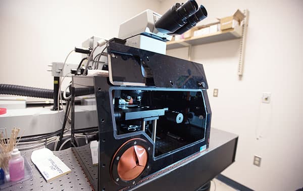
The Olympus FV1000 MPe Multiphoton and laser scanning confocal microscope is based on an Olympus BX61wi upright microscope and is equipped with a Mai Tai HP tunable laser (690-1040 nm) and two non-descanned detectors (2 PMTs). Filters for these detectors are red/green, yellow/cyan and green/violet. A 25x (NA 1.05, WD 2 mm) objective for MPe work is available. For confocal work, 405nm, 488nm, 515nm, 559nm and 635 nm excitation lasers are available for sequential photoactivation and imaging. A SIM scan unit is available for simultaneous photostimulation with a 405 nm laser. Available confocal objectives are 10x (dry), 20x (dry), 40x (oil), 60x (water), 100x (oil) and 100x (water). Multiplane images possible. No stitching.
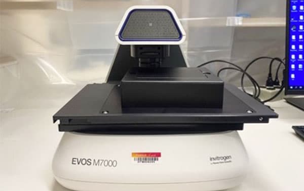
EVOS M7000
The EVOS M7000 Imaging System is a high-performance, fully automated, inverted, multi-channel fluorescence and transmitted light imaging system. The EVOS M7000 Imaging System offers these advantages: outstanding image quality and versatility with a five-position objective turret (2x, 4x, 10x, 20x, 40x), four-color LED fluorescence (available excitation/emission cubes: Texas red, CFP, YFP, DAPI, GFP), RGB color image collection, and 3.2 MP CMOS color and B/W cameras. The objectives are long working distance capable of imaging through plastic plates/dishes and glass slides. Exceptional usability with fully automated X/Y scanning stage, multi-position well scanning, autofocus, Z-stack, and tile-stitch options.
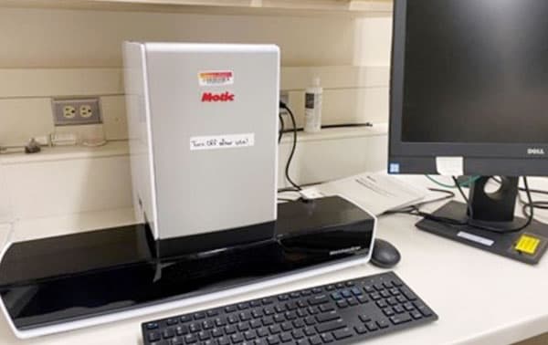
Motic EasyScan Pro6 Digital Slide Scanner
Upright microscope with 10x and 20x objectives, as well as a zoom feature. Brightfield with RGB color camera only. Perfect for H&E-stained sections. Digitally scan up to 6 slides in a single pass.
Analysis Station – Nikon NIS-Elements
Nikon image analysis software at a computer workstation. Includes deconvolution, tracking, and AI modules.
Analysis Station – Slidebook
Image data, especially .sld files, can be analyzed by a wide variety of tools for image processing, including mathematical operations, statistics functions, analysis scripting and import/export to/from other microscopy software.
Analysis Station – Imaris East
Oxford Instruments' image analysis software. Modules include: WholeSlide Analysis Package, ImarisXT, Voloom, MeasurementPro and ImarisVantage. Workstation also has MathWorks' MatLab with Image Analysis package.
Analysis Station – Imaris West
Oxford Instruments' image analysis software. Modules include: MeasurementPro, ImarisTrack, ImarisColoc, FilamentTracer, and ImarisVantage.